


By Kelly Gravitt, DVM, DABVP (Equine)
A diagnosis for a horse’s lameness is determined by first localizing where the lameness is coming from and then utilizing imaging to find the reason for the lameness. Once the lameness is localized by watching the horse move, flexing the limbs to exacerbate the potential areas of injury and using local anesthetics to “block” or numb the source of pain, imaging is then performed. Diagnostic imaging allows veterinarians to diagnose and monitor treatment of different causes of lameness and is an invaluable resource in lameness examinations.
Digital radiography, or x-rays, are one of the most common imaging modalities used when diagnosing lameness. Radiographs help veterinarians visualize bony structures inside the horse’s body and can diagnose diseases such as arthritis, osteochondritis dissecans (OCD) and laminitis. With advances in technology, radiograph units have become much more accessible to our equine patients. The Mizzou Equine Field Service utilizes a portable radiograph unit that does not require an electrical outlet. This can be especially helpful in situations of severe lameness when a horse is unable to move. The radiograph equipment can go to the horse’s location in these scenarios, decreasing the risk of further discomfort or injury.
If a horse is thought to have a soft tissue injury, a veterinarian may use a digital ultrasound for imaging. Ultrasonography uses sound waves to detect different densities and create an image of the structure being examined. Bone appears as white, fluid as black, and all the other structures appear as shades of gray in between. Ultrasonography can detect lesions in tendons or ligaments and can even detect some bony defects two weeks sooner than radiographs. The Mizzou Equine Field Service has a portable ultrasound machine that allows for ease of use on the farm. In addition to diagnosis of lameness, digital radiography or ultrasonography can also be used in treatment procedures, such as joint injections.
In cases that need more advanced imaging, the Mizzou Equine Clinic offers the ability to perform a CT scan or MRI.
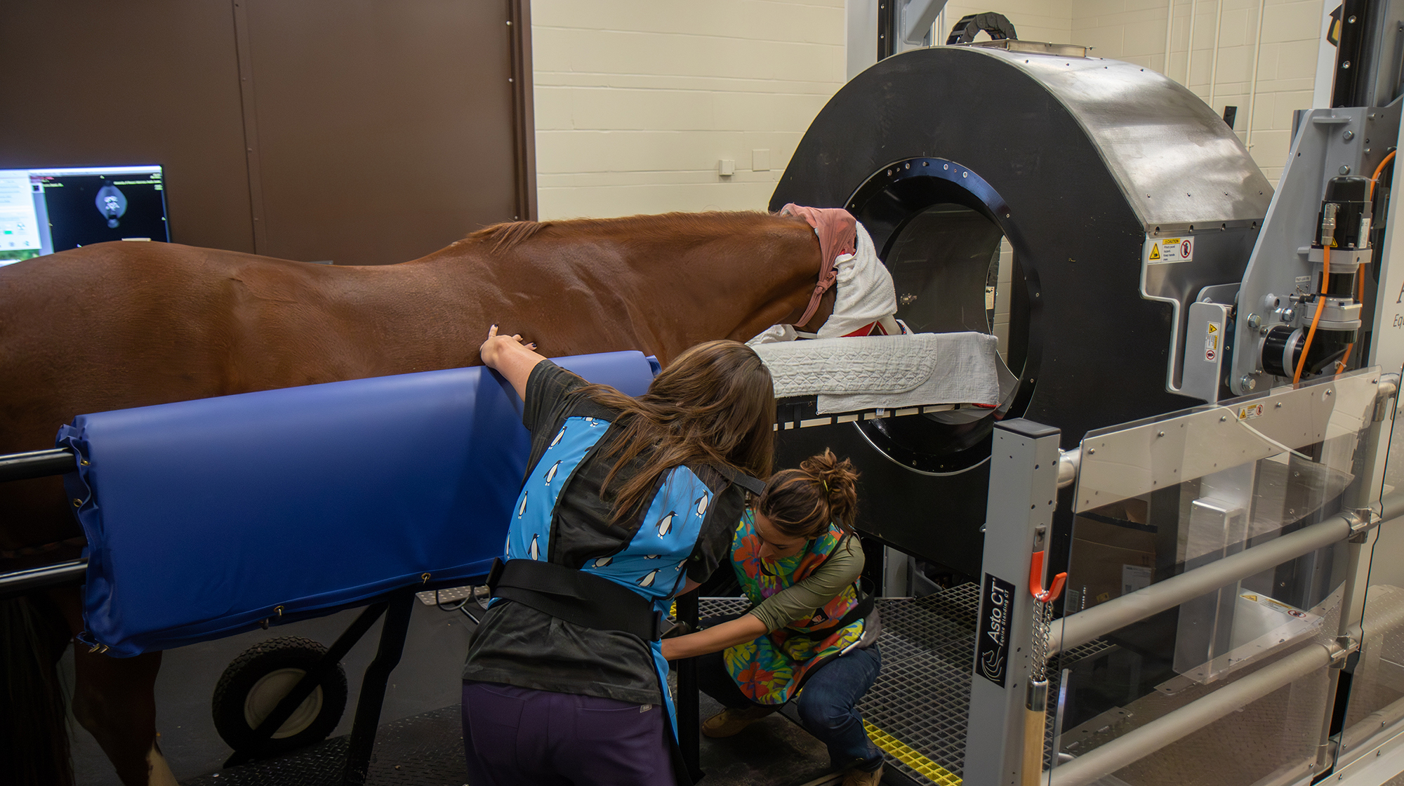
Computed tomography (CT) scans provide veterinarians with high-resolution 3D images to diagnose complex conditions of the horse. The Mizzou Equine Clinic offers a standing CT which allows for imaging of the head, parts of the neck, and limbs to the level of the knee or hock. The standing CT only requires light sedation and allows for high-quality images with 3D image reconstruction. This imaging modality is especially helpful for bony injuries and can provide earlier detection than radiographic imaging alone.
Magnetic resonance imaging (MRI) uses magnetic fields to create high-quality images of a horse’s bones, joints and soft tissues. This modality is often utilized in cases of complex lameness issues, especially those that originate in the horse’s hoof capsule. This is because an MRI allows for deep tissue insight that other modes cannot provide. An MRI may be indicated when a diagnosis has not been found by other imaging means or when very detailed images of both the soft tissue and bone are needed.
Diagnostic imaging is a vital part of lameness diagnosis. It allows for a veterinarian to determine the cause of lameness, assess the severity of the lesion and formulate a plan for treatment. Imaging also allows for a veterinarian to monitor the healing process of an injury or the progression of disease. If your horse is experiencing lameness, or has a history of lameness, talk to your veterinarian about an examination and your options for imaging.
