


Today’s world is fast-paced. We fuel ourselves with microwave meals at home and fast food on the go. We want high-speed internet, next-day delivery and eyeglasses in an hour. We expect fast weight loss from fad diets and immediate results from the latest training regimen.
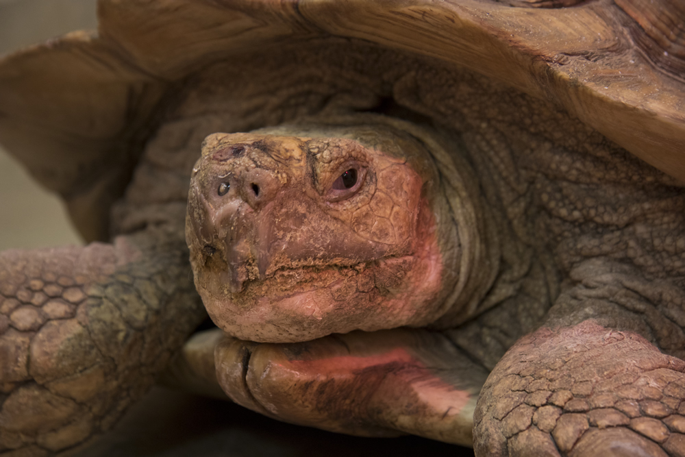
Mr. Pibb, a 60-pound sulcata tortoise, was referred the Veterinary Health Center for treatment of a large hole between the oral and nasal cavities.
Yet, Aesop’s fable reminds us that the slow and steady tortoise wins the race. Sometimes, by a nose.
Mr. Pibb, a 60-pound sulcata tortoise, had a nose that was not a winner. It was a problem.
“Mr. Pibb’s owner called us initially,” says Meagan Brophy, DVM, a clinical instructor at MU’s Veterinary Health Center (VHC). “He is in southwest Missouri and is a very educated owner.”
“Dr. Penning, Mr. Pibb’s owner, is an assistant professor in the Department of Biology and Environmental Health at Missouri Southern State University in Joplin,” adds Jennifer Blakely, a veterinary student who assisted with Mr. Pibb’s treatment.
“Both the owner and his referring DVM are very well-educated on this sulcata tortoise,” Brophy says. “I think they are friends outside of their professional relationship, and the DVM has 30 sulcata tortoises himself, so he is also very comfortable with the species — unlike us.”
“The owner called us and said, ‘My tortoise has an oronasal fistula,’” Brophy recalls. “The DVM had already anesthetized the tortoise and flushed out the area in question and said, ‘I don’t think there’s anything I can do for you. You should try calling the vet school at MU.’”
An oronasal fistula is a hole between the oral and nasal cavities.
“This is something that we commonly see in dogs, and it can be challenging to close these areas, Brophy says. “Our poster children for this condition are older dachshunds, dogs that have long, skinny snouts and large canine teeth. They can get infection and periodontal disease in those canine teeth and it starts to erode away the bone between the oral and nasal cavity. If you have an older dachshund on the table, and it has a canine that looks bad, you can flip a coin on whether it’s going into the nasal cavity. That’s how common it can be with dachshunds.
“So, when we got a phone call about this tortoise saying oronasal fistula, our brain pops into that dachshund mode — we fix these all the time,” Brophy says. “We don’t know anything about tortoises, but we’ll try.”
Brophy says she was upfront with the owner, telling him “We don’t know anything about tortoises, but we know people who do, so if you’re willing to work with us, we’re willing to work and learn about these guys and try to help him as much as possible.”
“We didn’t see the patient until the morning of the procedure. The owner sent us some pictures that were intra-operational, showing the referring DVM flushing out the area, which helped to show us the extent of the wound, or fistula, and its outward appearance, because it was malformed.
“In one picture the owner sent, some of the tissue looked devitalized on the top of his little snout. It almost looked like something was coming out of his nose, like a piece of food or something,” Brophy says. “The owner said, ‘Well, we knew something was up when we saw the swelling, and then we picked some food out of his nose. Then, we picked a pumpkin seed out of his nose. It was July, and he hasn’t had pumpkin seeds since fall. So, the food and whatever was causing that impaction had been up there since, at least, last fall.’”
Brophy says her theory is that fibrous material somehow punctured the palate of the tortoise and created an infection that festered. “They don’t have teeth on top; it’s just a beak and all this fibrous tissue. That’s what made it super challenging.
“Going back to our dachshunds, the treatment is to remove the tooth — remove the igniting cause of the periodontal disease — and close up the gum tissue. Dog gum tissue — mucosa is the proper term for it — is stretchy, much like human tissue. We can manipulate it, so it can close. We can create a false separation between the oral and the nasal cavities in a dog, and that’s how we fix it, by recreating that separation. But, with the tortoise, we’re not dealing with our dachshund’s stretchy mucosa. It is just bone and fibrous tissue. There was nothing we could have done that’s remotely like what we would do on a dog. We did not really know that until we got into the procedure that morning.”
To understand the extent of Mr. Pibb’s condition, Brophy ordered a computed tomography scan.
“Our main reason behind the CT was to determine whether cancer is causing this problem, or if it’s just a festering trauma that has gone out of control. The owner had said, ‘If you see something that looks like neoplasia or something similar, call me and I’ll come say goodbye.’”

A CT scan showed the extent of the damage to Mr. Pibb’s oral and nasal cavities, but showed no indication of cancer.
The CT showed that the void went extremely deep, to where there were hardly any normal nasal turbinates on that side, Brophy says. “It went from his little nose all the way to the back of his throat, at least 3 inches deep and 1.5 inches wide. It was massive, incredibly extensive, so we moved him up to our dental suite. The first thing we did was culture the wound and then we started to remove some devitalized tissue from the area.”
“We used a punch biopsy tool and removed a circle of necrotic tissue leaving an opening on the top of his nose into his sinus cavity,” Blakely explains.
“Once we did that on the dorsal aspect of the area, we actually found still more debris and food stuck up there after the flushing,” Brophy says. “We knew that had been there for quite some time because the owner said he had changed Mr. Pibb’s diet the past few weeks, trying to not get so much debris up in that hole while we were waiting and preparing for this day.”
Mr. Pibb had been eating a pelleted turtle-specific diet. “The material we discovered in the defect was more consistent with roughage or plant-like material,” Blakely says. “There was also a large amount of purulent and mucoid material as well as some remnants of a pelleted food substance.”
“It was in that nasal cavity and all the way back down into the throat,” Brophy says. “We had to flush it really well multiple times, alternating with suction to get that out of there, because it was pocketing. That was showing up on CT.
“It wasn’t in the initial plan to close that huge void on that day,” Brophy says. “We were trying to really limit our anesthetic time, assess and rule out neoplasia, take a culture and maybe send the patient home with some antibiotics. Maybe we could get the tissue a little bit healthier, and then have him come back and we would try to close it.
“Once we got in there, we saw there was no way we could close it, even if the tissue was healthy,” Brophy says. “The bone was gone. We would never be able to stretch that tissue enough to close the area. Plus, the deformity had been there for months and months, so the body thought it was finished healing. There is just no way you’re going to bridge a 1.5-inch gap in a sulcate tortoise.”
*****
How do you fix a 1.5-by-3-inch gap inside the head of a tortoise? Do some horsing around with it. Brophy consulted with Joanne Kramer, an associate teaching professor of equine surgery at the VHC.
“She suggested using a two-part material they mix up and put in special boots to treat horses with laminitis. When you mix it, it changes from a putty to something the consistency of a Super Ball: really rubbery and bouncy.
“I had never worked with it before, so that morning I was experimenting to get the consistency I wanted, in the time I wanted,” Brophy says. “I thought I had it straight. I didn’t.
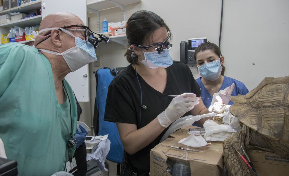
Richard Meadows, DVM, DABVP, Curators Teaching Professor, Meagan Brophy, DVM, clinical instructor, and then-third-year veterinary student Jenny Blakely, work to clean out and repair the gap in Mr. Pibb’s nose.
“To plug that hole, I had access from the top and from the mouth,” Brophy continues. “I was trying to make it a C-shape, where it would attach to the bone around the hole and the palate, to give it the strength of something like a ledge. He could still chew on it but it would be so dense that nothing he would eat would penetrate it. I wanted to create a false palate, where we would still have access to the nasal cavity to form this hole.
“I thought I had perfected the consistency, so I made it pretty, but it never cured,” Brophy says. “I checked on the patient in anesthetic recovery, but the material still had not cured. This tortoise was probably about to wake up. I pulled that first attempt out — the one I had probably spent 30 minutes trying to make pretty — mixed another batch and shoved it into the void space in about five minutes. This patient wasn’t even on anesthetic gas; it was in recovery, mildly sedated. We put a passive catheter through the nasal passage, to make sure we didn’t block it off too much, so he could still breathe through his nose.
“We made sure all of that was good, and then we sent the owner home with some injectable antibiotics and some sterile saline with instructions to flush through the hole on the top of the nasal cavity once a day,” Brophy says. “We needed to keep that area open and make sure we were not making a perfect environment for anaerobic bacteria.
“Mr. Pibb recovered very well, thanks to our anesthetic department,” Brophy says. “They did a wonderful job in researching and anesthetizing the animal, and totally rolling with the punches, going all the way to Plan C by the time they got him anesthetized.”
“The patient was here at 7:30 a.m. and gone by 2:30 p.m.,” Blakely says. “Some of our canine and feline patients don’t leave the hospital until 5, so this was a wonderful recovery. It was a really good experience to work with Dr. Brophy, Dr. Richard Meadows and all the other doctors who played a role in Mr. Pibb’s experience. This was not something we see every day. In veterinary medicine as a whole, I think you need to be able to make parallels between your patients, regardless of the species, and learn from each experience. I plan to go back to rural Missouri to work in a mixed-animal practice. I hope to be able to implement some of the things I learned from Mr. Pibb into my future experiences. However, if I ever come across another tortoise, I’ll know what to do.”
To clarify, tortoises are land dwellers with round, stumpy feet. Turtles spend most of their time in water; they have webbed feet for swimming and flatter backs. Terrapins spend time both on land and in water. Family members survive in almost every type of climate and are found on every continent except Antarctica. Of the approximately 300 species, 129 are vulnerable, endangered or critically endangered.
Testudines cannot “come out of their shell.” A tortoise’s shell is part of its skeleton, not just an exterior shield. Made up of more than 50 bones, including its rib cage and spine, the shell grows with the animal.
Sulcata tortoises, like Mr. Pibb, are the largest mainland tortoise. Sulcatas easily reach 30 inches in length and well over 100 pounds in heft. Some males even reach 200 pounds, but those examples would be in the upper age range.
Sulcatas are also known as African spurred, African spur thigh or simply spurred tortoise. Originating in north central Africa, sulcatas have shown an amazing ability to adapt to various climates and habitats in captivity. Sulcata tortoises are bred on a large scale in the United States, generally in the Southern states from coast to coast, where it is easier to keep them outdoors year-round. Their low cost, lively personalities and curious intelligence make them popular with first-time tortoise owners.
The origin of turtles has been one of the last unanswered questions in vertebrate evolution. Traditionally, paleontological and morphological studies placed Testudinidae as evolving either from the ancestor of all reptiles, or as evolving from the ancestor of snakes, lizards, and tuataras. Recent genetic studies indicate that Testudinidae are related more closely to dinosaurs, crocodilians and birds than to snakes and lizards.
