


What is Pulmonary Hypertension?
The cardiopulmonary (heart and lungs) system is an intricate system that pumps blood, which carries oxygen, to all of the parts of the body. The system relies on varying pressures generated by the heart and blood vessels to control blood flow. After the blood has dropped off its oxygen load to the tissues, it returns via the veins to the right side of the heart. This deoxygenated blood is then sent to the lungs to become oxygenated again. This oxygenated blood is then moved to the left side of the heart, which pumps the blood out through the arteries to the rest of the body. Because the oxygenated blood has farther to travel, the left side of the heart must pump harder (generate more pressure) in order to get the blood to the entirety of the body. The right side of the heart is a lower pressure system because it only has to pump blood to the nearby lungs. Pressures in blood vessels are measure in units of “millimeters of mercury,” or “mm Hg.”
Figure 1. Cardiopulmonary circulation. The right side of the heart pumps blood from the body to the lungs
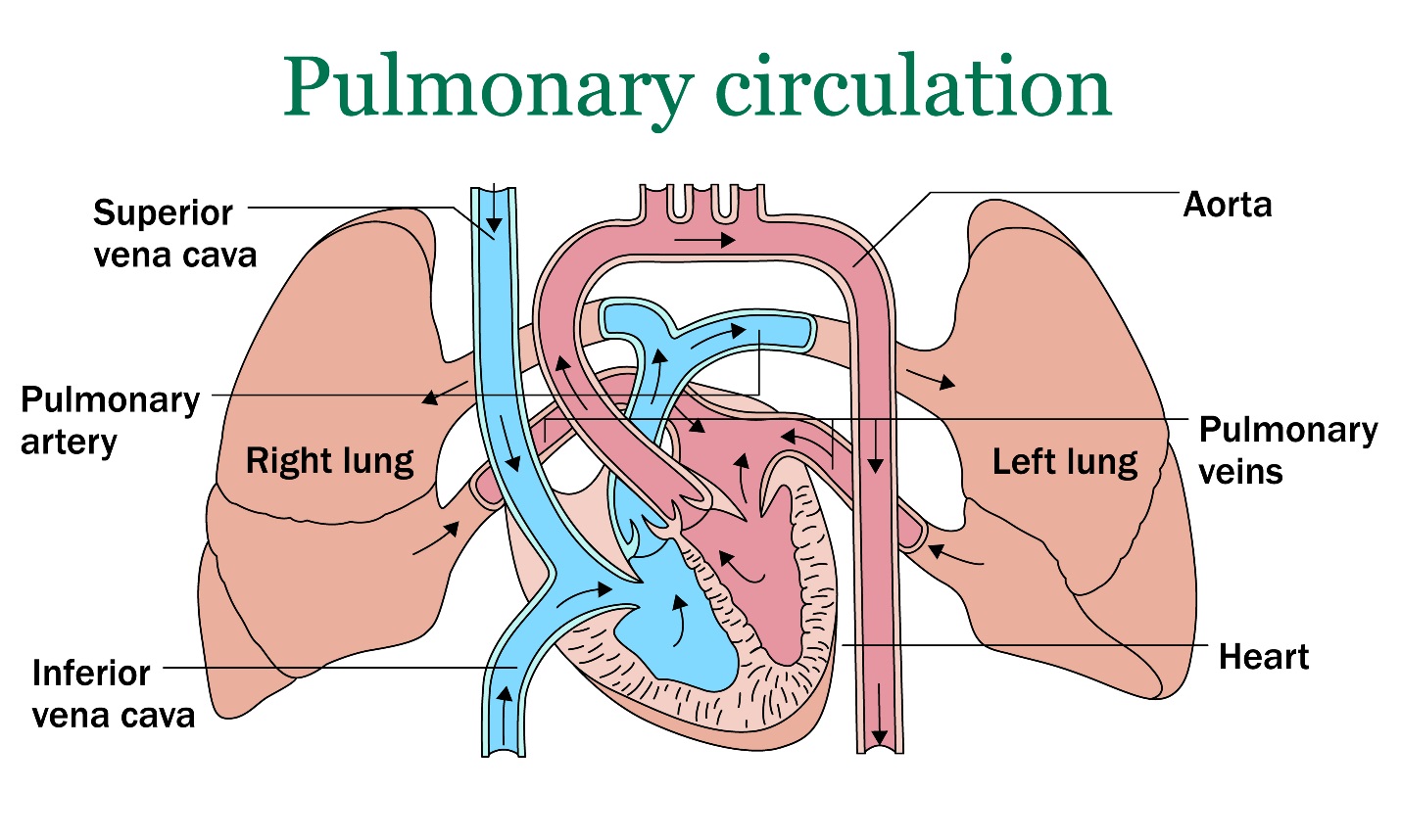
(blue), while the left side of the heart pumps blood from the lungs to the body (red).
Normal pressure in the lungs and pulmonary arteries (vessels from right side of heart to lungs) is 25 mm Hg systolic (when the heart pumps) and 10 mm Hg diastolic (when the heart relaxes and fills). Pulmonary hypertension is defined by a systolic pulmonary arterial pressure of >30 mm Hg. Pulmonary pressure can be elevated for a variety of reasons that can be divided into five classifications of pulmonary hypertension, as shown in the table.
|
Classifications of Pulmonary Hypertension |
Specific causes |
|
Pulmonary arterial hypertension |
Heartworm disease Congenital systemic-to-pulmonary shunts Atrial septal defect (ASD), ventricular septal defect (VSD), patent ductus arteriosus (PDA) Idiopathic Pulmonary veno-occlusive disease Necrotizing vasculitis/arteritis |
| Pulmonary hypertension with left heart disease (increased pulmonary venous pressure) |
Mitral Valve Disease Myocardial Disease Other Left-sided Heart Disease |
|
Pulmonary hypertension with pulmonary disease or hypoxia |
Chronic Airway Obstructive Disease Interstitial Lung Disease Neoplasia Obstructive sleep apnea High-altitude Disease Pulmonary edema–artery vasoconstriction |
|
Pulmonary hypertension due to thrombotic and/or embolic disease |
Heartworm Disease Thromboembolism Immune-mediated hemolytic anemia, neoplasia, protein losing disease, hyperadrenocorticism, Disseminated intravascular coagulation (DIC), sepsis, trauma, recent surgery |
| Miscellaneous | Compressive mass lesions |
Many cases of pulmonary hypertension in animals are related to left side heart disease. Degenerative valve disease is a very common disease seen in dogs, particularly older small breed dogs. The mitral heart valve, the valve between the two chambers (atrium and ventricle) on the left side of the heart, becomes thick and defective. When this valve begins to fail, pressure builds up in the left atrium and pulmonary veins, increasing the pressure in the lungs, leading to pulmonary hypertension.
Another common cause of pulmonary hypertension in dogs is due to respiratory disease. Disease in the lungs affects the ability of the vessels to dilate appropriately, increasing the resistance in the vessels. This increased resistance results in increased pressure in the pulmonary vasculature. Increased pressure in the pulmonary vessels causes fluid to “back up” into the right side of the heart, forcing the right side of the heart to pump harder to push blood to the lungs. This can eventually lead to failure of the right side of the heart.
Heartworm disease and pulmonary thromboembolism (blood clots in the lungs) are also important causes of pulmonary hypertension in dogs, and occasionally in cats too.
Since the heart and the lungs are so closely associated, high pressure in one system results in high pressure in the other. Pulmonary hypertension caused by lung disease can cause failure of the right side of the heart, while pulmonary hypertension caused by heart disease can cause lung dysfunction.
What do dogs with pulmonary hypertension look like?
Dogs with pulmonary hypertension can present to their veterinarian for a variety of reasons because pulmonary hypertension can be caused by many different disease processes. Common presentations are associated with poor heart or lung function. This can appear as lethargy, exercise intolerance, a cough, difficulty breathing, fainting episodes, or a fluid-filled stomach. Middle-aged to older small breed dogs are the most commonly affected with pulmonary hypertension; however, any breed dog can develop the disease.
How is pulmonary hypertension diagnosed?
- Right heart catheterization
- Right heart catheterization is considered the “gold standard” of diagnosis and is commonly used in human medicine. However, it involves placing a long wire inside the jugular vein at the level of the neck, extending down until it enters the right atrium of the heart, the right ventricle, and then the pulmonary artery. This is invasive and therefore not commonly performed in veterinary medicine. Catheterization of the heart allows for the most accurate measurements of pressure.
- Echocardiography
- Echocardiography, ultrasound of the heart, is the most common way to diagnose pulmonary hypertension in veterinary medicine. Echocardiography can measure blood flow moving across the tricuspid valve (between the right atrium and ventricle) and pulmonary valve (between the right ventricle and pulmonary artery). Additionally, echocardiography can visualized thrombi (blood clots) or heartworms that could be the cause of pulmonary hypertension.
How do we treat pulmonary hypertension?
Treatment of pulmonary hypertension is ideally centered around treatment of the underlying cause of the hypertension. For example, heartworm infection must be treated, or disorders causing blood clots should be addressed. Unfortunately, not all causes of pulmonary hypertension can be fixed.
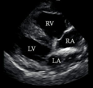
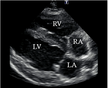
Images 1a (left) and 1b (right): Echocardiograms of a normal heart (a) and heart affected with primary pulmonary hypertension (b). RV = right ventricle. RA = Right atrium. LV = Left ventricle. LA = Left ventricle. In the affected heart, the right side of the heart is enlarged due pulmonary hypertension. In a normal heart (1a), the left side of the heart is larger because it pumps blood to the entirety of the body.
Images captured by Kelly Wiggen, DVM, Cardiology Resident, University of Missouri
Additional treatment includes medications that cause dilation of the pulmonary vessels. Dilation of the vessels decreases resistance within the pulmonary vessels and therefore decreases the pressure. These medications target different pathways involved in pulmonary vasodilation including endothelin, prostanoid, and nitric oxide pathways. The two drugs used most commonly to treat pulmonary hypertension are sildenafil (also known as Viagra) or tadalafil (also known as Cialis®). The drugs work by acting at the level of the vessels in the lungs, causing dilation and reducing pressure build up. Sildenafil (Viagra) was a drug originally produced for pulmonary hypertension in humans, however the drug was found to profoundly affect other areas of the body, which is now what the drug is used for in humans. Interestingly, sildenafil can also cause vasodilation in the vessels of the ears in dogs which can cause their ears to stand up more than usual.
Prognosis
Prognosis is highly variable based on the underlying cause of pulmonary hypertension. Addition of sildenafil and tidalafil has not necessarily been associated with an increased survival time, but has shown to increase quality of life and exercise capacity. Monitoring of pulmonary hypertension is performed by echocardiogram and frequency generally depends on severity of hypertension, administration of medication, and cause of pulmonary hypertension.
Written by Emily Soltis, Class of 2019
