Fluoroscopy is a type of advanced imaging that allows us to view the inside of the body while it is in motion. The image is similar to that of a radiograph, or x-ray, but instead of viewing a single still image, many images are produced by fluoroscopy to capture motion in real time. Essentially, this produces an x-ray movie that we can watch and replay. This moving image is helpful to view both the structure and function of organs to identify abnormalities that may not be apparent on a traditional x-ray that shows a single snapshot in time.
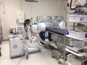
 The fluoroscopy machine at the Veterinary Health Center includes a main body unit that is connected to a C-arm unit, which extends around where the dog or cat will be positioned on the table.
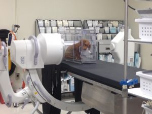
A patient undergoing fluoroscopic imaging will sit or stand on the table inside a plastic box. The box is positioned within the two sides of the C-arm. The machine generates a continuous x-ray beam that is targeted at the dog or cat.
Animals must hold perfectly still for x-rays, but this is not so for fluoroscopy. While in the box on the table, the animal will be able to sit, stand or lie down. They will likely be coaxed to do different things during the examination, in order to capture images in different positions. The main body unit and the C-arm move together alongside the animal to adjust to the height and location on the animal, based on size and position.
We often use fluoroscopy to visualize body systems that move, including the heart, the respiratory system (airways), and digestive system. Using fluoroscopy, we can appreciate the motion that occurs while an animal is swallowing and breathing. This allows us to see when abnormalities occur in order to diagnose and monitor various disorders. We can watch food as it moves from the mouth, to the esophagus (food pipe) and into the stomach. We can also watch how the trachea (wind pipe) changes shape as air passes through.
Swallow Study Using Fluoroscopy
Your veterinarian may have recommended a fluoroscopy swallow study for your pet. Common reasons to perform a swallow study include, but are not limited to:
- Difficulty swallowing food
- Suspected motility disorder of the esophagus
- Signs of regurgitation (spitting up the food after swallowing)
When a swallow study is performed, your pet will be placed in the box as described and pictured above. Several different types of food mixtures with different consistencies will be prepared ― liquid, semi-formed soft food, dry kibble. Each mixture will contain a contrast material that shows up on the fluoroscopic exam very well. When the food has contrast mixed in with it, we can more easily see the food and watch it travel down the esophagus. While your pet is swallowing each food mixture, we evaluate the motility of the parts of the body associated with this process. Read below for brief descriptions of a few disorders was can observe through this process.
Gastroesophageal Reflux Disease (GERD)
After food is swallowed, it travels down the esophagus and into the stomach. In the stomach, food is broken down into very tiny pieces so that it can be absorbed to provide nutrients. The environment of the stomach is specialized so that the stomach is able to perform this function. More specifically, the environment is acidic. The esophagus is not meant to hold acid like the stomach. There is a tight muscular ring separating the esophagus from the stomach. This ring is only supposed to open when stimulated to allow food to pass into the stomach but should then close to keep acid from the stomach out of the esophagus.
In animals with gastroesophageal reflux disease (GERD), contents from the stomach travel backward into the esophagus. On fluoroscopy, we can often watch reflux occur. Some animals will regurgitate this material, or bring it all of the way into their mouths. This can be dangerous. The acidic and other digestive contents of the stomach cause irritation to the esophagus; we suspect that this feels somewhat like heartburn that you may experience. Some animals with GERD lose their appetite. If this continues for a long period of time, more serious complications and damage to the esophagus may occur. If an animal is regurgitating this material, there is a risk that it will be inhaled into their lungs, or aspirated, causing pneumonia. GERD can also lead to other non-GI conditions including diseases of the airways. In fact, GERD is one of the leading causes of chronic cough in humans.
This video shows gastroesophageal reflux occurring in a dog. The dog is eating food material that contains the contrast, so it shows up well on the screen. She initially swallows the food and it travels down her esophagus toward the stomach. Then suddenly the food is forced to reverse and moves back up the esophagus toward the mouth. When this occurs, some material from the stomach enters the esophagus.
Esophageal Motility Disorders
Animals with difficulty swallowing food may suffer from a disorder in the motility of the esophagus. The esophagus is muscular and the muscles must contract to efficiently push food down into the stomach. In addition, there are two muscular rings that food must pass through ― one is located at the top of the esophagus, near the mouth, and the other is located at the bottom of the esophagus, where the food passes into the stomach. Motion of these muscles is well coordinated so the movement of food is rapid. If the muscles in the esophagus or either muscular ring are not able to contract properly, then it cannot carry the food down the pipe and into the stomach as it should. This means that food may move down the esophagus slower than it should, or that it may become stuck there for a period of time. Animals with these types of disorders may cough or spit up their food as a result, putting them at a higher risk of breathing food into their lungs. We can observe these conditions with fluoroscopic examination, allowing appropriate treatment to be recommended.
This video shows an esophageal motility disorder in a dog. The dog is eating food material that contains iohexol contrast, so that it shows up well on the screen. After she swallows the food, it remains in the esophagus and collects there. The food should pass through quickly and enter the stomach. Instead, it accumulates in the esophagus and does not enter the stomach. In this case, the muscular ring located at the bottom of the stomach is malfunctioning and is not opening up to allow food to pass through and into the stomach.
Dynamic Airway Examination
Your veterinarian may have recommended fluoroscopy due to concerns for a possible abnormality in the airways. If your pet makes loud noises when breathing, has difficulty breathing, or coughs frequently, there may be an obstruction in the airways. Disorders was can observe through this process include:
- Collapsing trachea
The trachea is the tube that carries air from the nose and mouth to the lungs, and back from the lungs to the nose and mouth, during normal breathing (inspiration and expiration). It is supported by very firm rings that are made of cartilage that help it keep its tubular shape. In some animals, this cartilage becomes weak and is not able to keep the trachea completely open all of the time. When this occurs, the trachea collapses, triggering the animal to cough. Some animals have difficulty breathing when the trachea collapses. Sometimes, this can be visualized on a radiograph, but this can be difficult to capture on a single x-ray picture because the trachea does not remain closed all of the time. On fluoroscopy, we can watch the trachea collapsing as the animal is breathing or during cough. We can use this to determine the degree of collapse as well as the location in the trachea where the collapse is occurring. Based on this information, appropriate treatment recommendations can be made. Sometimes, treatment will include placement of a tracheal stent. This too is done while watching with fluoroscopy.
This video shows a dog breathing in and out. It focuses on the dog’s chest and lungs. The lungs, located in the chest area, expand when the dog breathes in and become smaller when the dog breathes out. Notice the white tubular structure going down the neck that leads into the lungs. This is the trachea. Look closely at how the width of the trachea changes as the dog breathes in and out. The area that is further down the neck and closer to the lungs best demonstrates the change. When the dog breathes in, the trachea is wider. When the dog breathes out, the trachea becomes narrower. This is abnormal. The trachea should stay the same size to allow good air flow. When it collapses, as in this video, the dog may cough or have difficulty breathing. At the end of the clip, we see the dog coughing. As she coughs, the trachea becomes even narrower, making it more difficult for her to breathe.
- Epiglottic abnormalities
- The epiglottis is located at the back of the throat. It is located between the trachea and esophagus. The epiglottis opens or closes so air can flow in and out of the lungs while breathing, but food does not go down the wrong way. When eating, the epiglottis flaps up to block the trachea so that food is directed down the esophagus. This function is important to ensure that food ends up in the stomach, not the lungs. An abnormally functioning epiglottis may cause an animal’s airways to become partially or fully blocked, leading to difficult or noisy breathing. If your pet experiences these signs, we can use fluoroscopy to watch the epiglottis to look for an abnormality it its motion.
If you believe your pet might benefit from a fluoroscopic study, please talk to your veterinarian about referral or call 573-882-7821 to schedule an appointment with the Small Animal Internal Medicine Service. It is important to hold your pet off of food the morning prior to the appointment.
Written by: Anna Delabar
Edited by: Leah Cohn and Megan Grobman


