


Veterinarians at the MU Veterinary Health Center diagnosed Maggie, a 2-week-old paint filly, with bilateral pleuropneumonia, a severe infection that is uncommon in foals.
At two weeks old, Maggie seemed happy and healthy. Then the foal suddenly began struggling to breathe, developed nasal discharge and a fever, and became lethargic. Her trainer, Owen Parker of Oak Grove, Mo., took her to his veterinarian, who referred the paint filly to the MU Veterinary Health Center for diagnosis and treatment.
Maggie, who is training to be a show horse, arrived at the VHC in the middle of the night. Veterinarians quickly performed a physical examination, routine blood tests and an ultrasound scan of the chest. The results led to a diagnosis of bilateral pleuropneumonia. Uncommon in foals, the severe infection primarily affected the pleural cavity, the surface of the lungs, the interior of the chest wall and the space between them. Normally, a small quantity of fluid in this space helps to lubricate the movement of the lungs against the chest wall. However, Maggie’s infection caused inflammation that provoked an excessive quantity of fluid to accumulate, which prevented her lungs from expanding completely and led to difficulty with breathing.
The initial treatment involved draining fluid from the chest, antibiotics and efforts to determine the cause. Determining the cause was important to ensure the infection wasn’t contagious and to identify the best antibiotics to use, said Dr. Philip Johnson, a professor of equine internal medicine at the MU College of Veterinary Medicine who treated Maggie.
In her chest fluid, veterinarians identified excessive fibrin, a thick inflammatory protein that interfered with their ability to drain the fluid and provide Maggie relief. The protein eventually forms a fibrous, or scar-like, attachment between the lung surface and chest wall, an outcome that would have permanently interfered with Maggie’s breathing.
To address the fibrin, veterinarians tried a novel treatment. They injected tissue plasminogen activator (TPA), a “clot buster” used in human medicine to treat strokes. When injected into the chest cavity, it can help dissolve fibrin.
“The more conventional treatment is drainage of chest fluid alongside antimicrobials, but fibrin accumulation, if it is significant, can both impede treatment effectiveness and render the patient incapable of a normal life from a breathing perspective,” Johnson said.
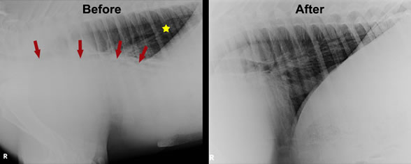
Left: Radiographic appearance of Maggie’s chest before treatment. The dorso-caudal lung fields appear normal (yellow star). Excessive fluid in the ventral part of the chest was evident (below the arrows). Right: Radiographic appearance of the foal’s chest after treatment. Excessive fluid in the ventral part of the chest is no longer evident, and the lung fields appear clear.

Left: Ultrasonographic image of the left pleural cavity obtained following draining of fluid. Notice the thick layer of fibrin (yellow double arrowheads) lining the chest wall and another thick layer of fibrin (blue double arrowheads) covering the surface of the lung (white double arrowhead depicts the wall of the foal’s chest). Residual pleural fluid is present between the two layers of fibrin. Right: Post-treatment ultrasonographic image of the ventral border of the foal’s left lung (arrows) showing absence of excessive pleural fluid and resolution of adherent fibrin.
Maggie improved quickly with the TPA, and both the fibrin and the excessive pleural fluid disappeared within a few days.

The left side of Maggie’s chest is drained, a process called pleurocentesis. The team caring for Maggie included Drs. Philip Johnson, Alicia Foley and Jamie Zimmerman; technician Jane Ebben; and veterinary student Nicole Freeman.
Maggie’s tests identified two pathogens that likely contributed to development of pneumonia, Streptococcus pneumoniae and a gammaherpesvirus. Although her veterinarians couldn’t be completely certain, they speculated that the gammaherpesvirus may have been the “first attack,” causing damage and inhibiting the foal’s immune system, Johnson said. They believe it was followed by the Streptococcus pneumoniae. She likely acquired the pathogens from other horses in her environment.
“Young foals have not developed a fully effective immune system,” Johnson said. “At this age they are encountering lots of potential respiratory viral and bacterial micropathogens from co-mingled horse stock.”
Maggie is now back to normal. Parker said he was very pleased by her care at the VHC, where he has had several horses treated. He said he has had great experiences with not just the veterinarians but also the staff.
“Our first pick is always the University of Missouri,” Parker said. “The staff are not like a lot of other places. You feel like a person there.”
|
