What are cataracts?
Cataracts are a common cause of blindness in dogs. A cataract is an opacity of the lens within the eye. The lens plays an important role in focusing light on the retina. A cataract prevents the light from reaching the retina, thereby impairing vision.
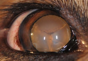
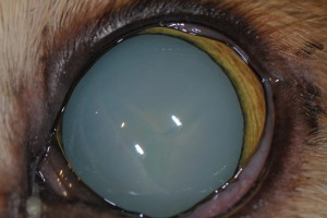
The amount of lens affected by the cataract can vary, with incipient cataracts occupying less than 10 percent of the lens (Figure 1), while 100 percent of the lens is affected when a cataract becomes mature (Figure 2).
Cataracts are also classified by age of onset; terms such as “juvenile cataract” or “senile cataract” are used by some veterinary ophthalmologists. Cataracts must be distinguished from a normal aging change in the lens known as lenticular or nuclear sclerosis, which causes a bluish appearance to the eye but typically does not cause blindness.
What causes cataracts in dogs?
The most common cause of cataracts in dogs is hereditary cataracts. Purebred dogs diagnosed with hereditary cataracts should not be bred. The second most common cause of cataracts in dogs is diabetes mellitus. Other causes of cataracts include advanced age, trauma, retinal degeneration, metabolic disorders or nutritional deficiencies. Depending on the cause of the cataract, it may not progress to a mature cataract.
How are cataracts treated?
Currently, surgical removal of the cataracts is the only means by which cataracts can be cured. Surgery is generally recommended when cataracts are causing vision impairment. Prior to performing surgery, a veterinary ophthalmologist will recommend performing both ocular and systemic diagnostic testing, including complete blood work, urinalysis, an ocular ultrasound and an electroretinogram, which tests the function of the retina.
Cataract surgery is performed under general anesthesia. Initially, a small incision is made in the cornea. Upon entering the eye, a circular hole (capsulorhexis) is made in the capsule of the lens. The cataract can now be accessed and removed with ultrasonic energy. Once all the cataract is removed, an artificial intraocular lens can be placed in the lens capsule. With the artificial lens, light can again be focused on the retina, and the dog’s vision will improve.
How successful is cataract surgery?
The success rate of uncomplicated cataract surgery with intraocular lens implantation in dogs is approximately 90 to 95 percent.
What are the risks and complications of cataract surgery?
Risks of cataract surgery include those due to anesthesia and those due to the surgery itself. The complication rate following cataract surgery is 5 to 10 percent. Some complications may require treatment with long-term medical management or even additional surgery.
Significant ocular complications include the following:
- Retinal detachment is a condition where the retina separates from the underlying tissue. This condition sometimes can be corrected with an additional surgery.
- Excessive inflammation, or uveitis, is often addressed by increasing the frequency of medication administration.
- Glaucoma refers to elevated intraocular pressure. Medical and surgical therapy can often control this complication. However, if the glaucoma is not controlled with these therapies, procedures such as an enucleation (removal of the eye) may need to be considered to restore patient comfort.
- Posterior capsular opacification, or “secondary cataract,” refers to the cloudiness of the lens capsule in which the artificial lens is implanted.
- Keratoconjunctivitis sicca, or dry eye disease, is a condition in which the lacrimal glands do not produce enough tears to maintain corneal health. This disease is typically managed with long-term medications. Dry eye disease is a common complication in diabetic patients.
What to expect after surgery
The patient is discharged the day after surgery wearing an Elizabethan collar (also known as an E-collar) to protect the eyes from self-trauma for approximately four weeks. The patient’s exercise should be limited during this recovery period. Topical medication will need to be administered four times daily for several weeks. The patient will also need to be re-evaluated by a veterinary ophthalmologist several times in the first year after surgery. Owners of dogs that have undergone cataract surgery commonly report a significant improvement in their pet’s quality of life.


