Cataracts are a leading cause of visual impairment among dogs. This opacification of the lens has various causes and may or may not progress to total blindness. The vision of affected dogs can often be significantly improved by surgical removal of the cataractous lens. Below are some important facts pertaining to cataract surgery and intraocular lens implantation in dogs.
What is a cataract?
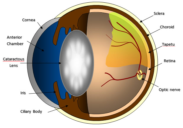
The term cataract refers to any opacity of the lens, a structure that lies within the eye (Figure 1). The function of the lens is to help focus light onto the retina which converts light energy into electrical impulses capable of being interpreted by the brain. Cataracts decrease vision by interfering with light reaching the retina. Advanced cataracts are a leading cause of blindness among dogs.

Figure 1. Diagram of the anatomy of the canine eye. A cataract, or white opacity in the lens, is present, which can impede vision and is a leading cause of blindness in dogs.
The majority of cataracts are the result of a genetic or inherited defect involving the lens. Several breeds of dogs are known to be predisposed to developing inherited cataracts. Dogs affected with inherited cataracts should not be involved in breeding programs. Cataract formation is less commonly associated with diabetes mellitus, advanced age, trauma, or retinal disease. Depending upon the cause, cataracts may or may not progress to total blindness.
The rate of progression is often variable, ranging from weeks to years depending upon the underlying cause of cataract formation. For example, cataracts associated with diabetes mellitus can develop quickly. In some cases, diabetic cataracts can progress very rapidly, requiring immediate attention to prevent or address secondary complications associated with the cataract progression.
A frequent consequence of cataract formation is the development of inflammation within the eye which, left untreated, can potentially damage the internal structures of the eye and lower the prognosis for a successful visual outcome following cataract surgery. Therefore, early evaluation by a veterinary ophthalmologist is recommended.
How are cataracts treated?
Presently, the only effective treatment of advanced or rapidly progressing cataracts is through surgical removal of the affected lens. This is accomplished under general anesthesia by making a surgical incision into the eye and using special instrumentation to ultrasonically fragment and remove the lens material. When possible, once the cataractous lens has been removed, an artificial intraocular lens is implanted into its place.
The success rate of uncomplicated cataract surgery is approximately 85-90 percent. This success rate may vary depending upon the overall health of the affected eye. An assessment should be made by a veterinary ophthalmologist to determine the relative risks and benefits of surgery.
It is imperative to note that even though there is a relatively high success rate, there are cases in which complications do arise. The consequences of these complications vary in severity and can include excessive inflammation, corneal edema (cloudiness), secondary glaucoma (an increase in the intraocular pressure), retinal detachment, intraocular infection, and total blindness. Some complications may necessitate intensive long-term medical therapy or even additional surgeries. Although uncommon, these complications do occur. Facts pertaining to the surgery will be discussed in detail during your dog’s initial cataract evaluation appointment.
Your local veterinarian’s role
Your local veterinarian plays a key role in proper maintenance of your dog’s overall health, and their role in the successful management of cataracts is no exception. Early detection of cataracts by your veterinarian with referral for subsequent evaluation by a veterinary ophthalmologist can have a positive influence on eventual outcome. Surgery is not indicated in every case; however, it often restores functional vision to dogs whose sight was impaired due to cataracts.
The cost of cataract surgery
Factors that determine the total cost include: whether one or both eyes will undergo surgery, the amount of presurgical workup required to screen for other diseases, and duration of hospitalization. Included in the cost is presurgical performance of an electroretinogram, a test to assess retinal function, and an ocular ultrasound, to screen for retinal detachment.
What to expect after surgery
Successful cataract surgery requires a firm commitment from the owner. Usually, the patient is admitted to the hospital the day before surgery and discharged the day after with restricted exercise, a special collar which protects the eyes from irritation for three weeks, and topical medications. Medical therapy will involve the instillation of various drops into the eyes multiple (four to six) times daily for the first few weeks after cataract surgery. Usually the medication is gradually decreased over the first month or so of therapy, depending upon your dog’s progress, but may be continued indefinitely. Multiple rechecks by our ophthalmology service will be recommended post-operatively.
Some post-operative inflammation and discomfort following cataract surgery is expected. Typically, the eyes may become more reddened than usual, there may be a slight increase in ocular discharge, mild corneal clouding, as well as varying degrees of squinting. All of these symptoms represent a normal response to surgery and should resolve over the first one to two weeks, with marked improvement noticed over the first three to four days. A common question after cataract removal is “how well can my dog see?” In certain cases, vision can be difficult to assess immediately after surgery but is usually noticeable within two days with continual improvement over the first two weeks. It is not normal for the eyes to remain painful or vision to either fail to improve or worsen over the first two weeks.
Medical therapy: The overall success of cataract surgery is highly dependent upon close compliance with the prescribed medication schedule. Medication should be given at evenly spaced intervals during your waking hours. If two drops are to be given at the same time, allow at least five minutes between drops to allow maximal absorption of each medication.
E-collar: This collar should be left in place at all times for the first three weeks following surgery. It will help prevent your dog from injuring the eyes during the initial post-operative period.
Exercise: Restrict your dog’s exercise. It is advisable to keep your dog confined to a small room indoors with supervised short walks outside. Avoid leashes when possible, using a harness if necessary, to limit the amount of pressure on the neck which can elevate intraocular pressure. Do not bathe your dog or encourage roughhousing until told to do so.
Rechecks: The initial recheck appointment is scheduled for one week following cataract surgery. This exam is crucial in the management of cataract patients as important recommendations will be made based on post-operative progress. Additional recheck appointments will be scheduled as necessary but are typically recommended at three weeks, two months, four months, and eight months post-operatively, then semi-annually or annually thereafter.
Problems: In most cases, complications are minimal. However, in the event an ocular problem develops or if you have questions or concerns pertaining to post-operative management, please notify the Ophthalmology Service at the University of Missouri at 573-882-7821. Emergency services are available 24 hours a day, seven days a week.


