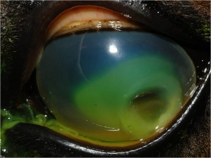
 The cornea is the clear external layer on the front of the eye and can be thought of as the “windshield” of the eye. A corneal ulcer occurs when a wound or scratch has occurred on the corneal surface. Although there are a variety of causes for corneal ulceration, most are caused by inadequate tear production, trauma or irritation from hairs touching the cornea. Hairs may contact the cornea due to eyelid conformational defects or congenital presence in locations on the face and eyelids where hair is not normally found.
A veterinarian is able to detect a corneal ulcer by staining the cornea with fluorescein stain (a green water-soluble dye) in the exam room. This fast test is required to correctly diagnose a corneal ulcer.
Once the cornea is ulcerated, the area can become infected by either bacteria or fungus, resulting in a more serious problem.
Treatment of corneal ulcers
Minor superficial ulcers can be treated successfully by your regular veterinarian and usually heal without complication. More serious ulcers will take longer to heal and often require assistance of a veterinary ophthalmology specialist to achieve complete healing. Sometimes advanced corneal ulcers will require surgery; this can range from a superficial debridement to a complete graft. During corneal healing, your pet will likely require an E-collar (cone) to prevent self-trauma.
Serious ulcers can cause permanent scarring of the cornea that results in vision loss, and early intervention in these cases is very important.
Signs of a corneal ulcer include pain, squinting, redness of the sclera (white part of the eye), tearing and discharge from the eye. In more serious ulcers, the cornea may appear cloudy or have a “divot” in one part, or there may be red blood vessels or pink tissue present on the corneal surface.


