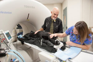
 Positron emission tomography (PET) and computed tomography (CT) is a unique offering of our radiology department. We are one of a limited number of veterinary facilities with the ability to perform PET/CT studies for animals within our hospital. Unlike radiographs, CT scans or MRI scans that show changes in anatomy, PET allows us to view physiological changes in the body and fuse these with a CT scan to provide anatomical mapping and changes as well.
We have a Canon Celesteion PET/CT scanner with a wide bore gantry. This system uses the newest technologies with a wide opening to allow for many sizes of animals. This allows us to perform a fast and sensitive scan with a small dose to the patient.
Our technologist is a certified nuclear medicine technologist with a specialty certification in PET imaging as well as CT.
 To perform a PET/CT scan, your pet is injected with a specific amount of a radioactive drug (radiopharmaceutical) designed to target the area of interest. A PET/CT scanner is used to generate an image from the radiopharmaceutical inside your animal to tell doctors about the function of that tissue. We also perform a CT to provide us with anatomical data to use with the PET scan. We then use this information to help in the diagnosis and treatment of the disease process.
Common tests our staff performs are glucose PET scans (FDG), bone scans (NaF) and thyroid scans (I124) as well as a variety of research studies utilizing our PET/CT scanner.
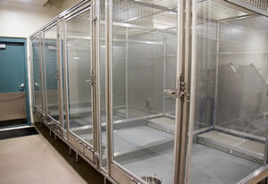
 We have a state-of-the-art isolation area to comfortably house our patients for a short time after the scan until it is safe for them to be released.


