There are two categories of disc disease. Hansen type I IVDH is a result of the nucleus pulposus (the jelly) being extruded and compressing the spinal cord. The majority of these occur in middle-aged chondrodystrophic breeds, such as dachshunds, beagles and basset hounds. Due to an inherited condition, the discs degenerate early in life.
Hansen type II IVDH is a result of a bulging of the annulus fibrosis (the outside of the donut) and occurs more commonly in older large-breed dogs. Intervertebral disc herniation can occur in the cervical spine and more commonly in the lower thoracic and lumbar spine.
Onset of clinical signs with Hansen type I IVDH is usually acute and progressive over time. The degree of clinical signs vary from back or neck pain only to paralysis. An animal with back pain may have a hunch-back appearance (kyphosis) and tense its abdominal muscles. The signs may be worse on one side because the disk is compressing that side of the spinal cord more than the other. Clinical signs can progress in order of functional loss: loss of proprioception (awareness of where the limbs are in space), loss of motor function (ability to move the legs), and loss of pain perception (inability to feel a toe pinch). Loss of bladder control is also a common occurrence in spinal cord injury.
Hansen type II IVDH in the thoracic and lumbar regions of the spine is slowly progressive and usually causes signs of hind-limb weakness (paraparesis), reluctance to rise or jump, and possibly back pain. Hansen type II IVDH also can cause neurologic signs that are asymmetrical due to lateralization of the disc herniation.
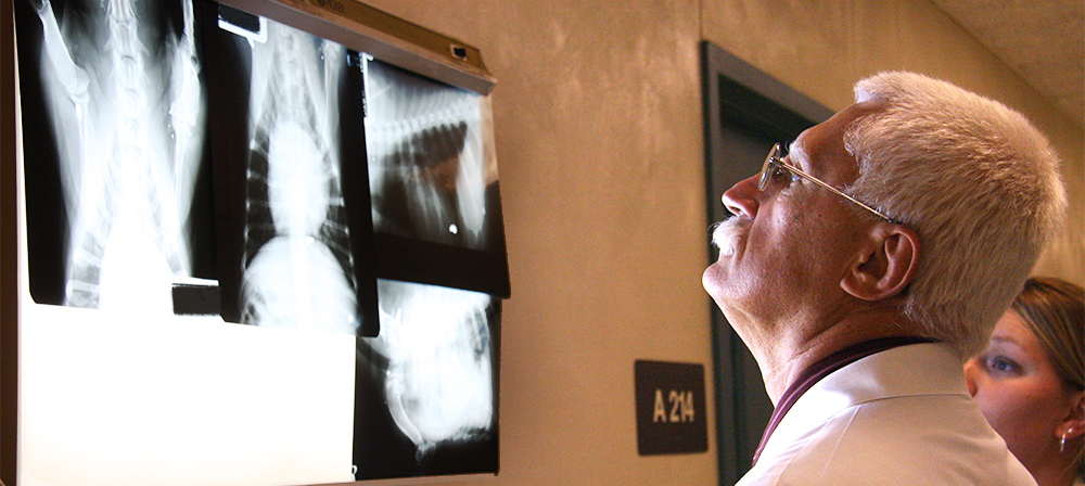
We may recommend spinal radiography (X-rays) before pursuing advanced imaging of the spinal cord. Spinal radiography enables visualization of mineralization in the intervertebral discs or narrowing of the intervertebral disk space. However, this technique will not enable us to visualize the damage to the spinal cord. Computed tomography (CT) or magnetic resonance imaging (MRI) of the spinal cord usually is required to diagnose spinal cord compression and damage. MRI is the gold standard, or superior, diagnostic test for intervertebral disk disease.
Conservative management may be recommended if the signs are mild and represent an initial episode. For example, if it is still able to walk and has spinal pain only, the dog may be a candidate for conservative management. Another indication for conservative management would be if the pet has other health problems that may preclude it from undergoing anesthesia and surgery. Most importantly, your dog requires strict confinement and restricted activity for six to eight weeks. This will require you to confine your dog in a small crate. This period of restriction gives time for the disc itself to heal. Pain management may also be necessary and usually includes anti-inflammatory drugs or opioids.
Surgery may be indicated if your pet has recurrence or progression of signs despite conservative management or if your dog is unable to walk at time of occurrence. There are a few different surgical procedures that can be performed to remove the portion of the disk that has herniated and is causing spinal cord compression. The neurosurgeon will make the decision which procedure is appropriate depending on the site of spinal cord compression. A hemilaminectomy is the most common surgical approach to gain access to the herniated material. Basically, the bone is removed on one side of the spine at the site of herniation. The herniated disc material is removed through this defect, which relieves the spinal cord compression.
After surgery, pain management, bladder management, kennel rest and physical rehabilitation are all important. Some pets may be unable to voluntarily urinate after surgery. Manual expression of the bladder can be performed by applying gentle pressure to the abdomen. A urethral catheter also can be passed into the bladder for emptying the bladder. If needed, there are drugs that can be administered to relax the sphincter and make bladder expression easier and more comfortable.
Strict kennel rest is important for six to eight weeks after surgery to give the surgical site adequate time to heal. Physical rehabilitation may also play an important role in the recovery process.


