Emergencies

Menu
  • About
      • About the VHC
      • Our Mission
      • Academic Department
      • Our Location
      • Veterinary Health Center – Wentzville
      • Contact Us
    • Personnel
      • Faculty Clinician Directory
      • Residents and Interns
      • Technical Staff
      • Staff
    • Close
  • Clients
      • Client Information
      • Make an Appointment
      • FAQs
      • Client Billing
      • Client Portal
      • Pay Your Bill Online
      • CareCredit
      • Pharmacy Services
      • Client Portal Instructions
      • Client Support
      • Client Satisfaction Survey
      • Touring the VHC
      • Lodging
      • Our Columbia Location
      • Columbia Resources: Ways to Refresh While You Wait
      • Wentzville Resources: Ways to Refresh While You Wait
      • Contact Us
    • Close
  • Small Animal

        • Hospital Overview


          573-882-7821
          After Hours: 573-882-4589
          Directions


          Contact Us

      • Anesthesiology
      • Cardiology
      • Community Practice
      • Dentistry
      • Emergency and Critical Care
      • Internal Medicine
      • Neurology/Neurosurgery
      • Nutrition
      • Oncology
      • Ophthalmology
      • Sports Medicine and Rehabilitation
      • Radiology
      • Reproduction (Theriogenology)
      • Shelter Medicine
      • Surgery
    • Close
  • Equine

        • Hospital Overview


          573-882-3513
          After Hours: 573-882-4589
          Directions

          Contact Us

      • Ambulatory Service
      • Anesthesiology
      • Internal Medicine
      • Lameness Service
      • Ophthalmology
      • Radiology
      • Reproduction (Theriogenology)
      • Surgical Services
    • Close
  • Food Animal

        • Hospital Overview


          573-882-6857
          After Hours: 573-882-4589
          Directions

          Contact Us

      • Hospital Care
      • Ambulatory Services
      • Anesthesiology
      • Ophthalmology
      • Radiology
      • Reproduction (Theriogenology)
    • Close
  • Clinical Trials
  • Referrals
      • Referring Veterinarian Information
      • The Referral Process
      • VHC Referral Form
      • VHC Wentzville Referral Form
      • Overnight Transfers
      • Veterinary Diagnostic Laboratory
      • RDVM Portal
      • Referring Veterinarian Satisfaction Survey
      • Faculty Clinician Directory
      • Referring Vets Guide (PDF)
      • Consultations
        • Cardiology Consultation Inquiry
        • Small Animal Internal Medicine Consultation Form
        • Orthopedic Referral and Estimate Inquiry
        • Neurology Referral Form
        • Nutrition Consultation Form
    • Close
  • Giving
  • Employment
  • Internal
You are here: Home / Small Animal Hospital / Oncology / Common Diagnostic Procedures
post

Common Diagnostic Procedures

onc3


Biopsy

Punch biopsy: A punch biopsy is a technique for removing a specific volume of tissue for examination using an instrument called a punch. Depending upon the size of the punch, a skin suture may be used to close the wound. The tissue is then placed in a formalin container and “fixed.” It can then be sent to a pathologist for histologic examination. It may be several days to a week before the pathology report is complete.

Tru-Cut biopsy: A Tru-Cut needle biopsy uses a much larger needle to obtain a small core piece of tissue. That tissue is “fixed” in formalin, processed for examination and then examined microscopically. This type of biopsy gives a larger specimen and preserves the architecture of the tissue, but it requires several days to get results.

Patients undergoing punch or Tru-Cut biopsies will generally require sedation or anesthesia depending on the tissue being biopsied.


Bone marrow aspirate

The bone marrow is the site where new blood cells are produced. Bone marrow aspiration may be used as a part of cancer screening or staging.

With the animal sedated, the area of the biopsy is numbed. The hair is clipped and the skin thoroughly cleaned. A special type of steel needle is inserted through the hard bone into the marrow (the pulpy material inside the hard bone). Some of this pulpy material is aspirated through the needle using a syringe. Sometimes, a piece of the marrow material is removed intact for biopsy. Although the results of an aspirate are typically available the same day, the core biopsy may require several days to evaluate.


Bone scintigraphy (scan)

A bone scan involves the use of a radioactive material injected intravenously to identify new areas of bone breakdown or growth. It is used to help diagnose conditions of the bone, including inflammation, cancer and metastasis to the bone. Patients must be housed in special radiation suites for 24 hours following injection of the radiotracer.


Computed tomography (CT) scan

Computed tomography uses a similar technique as is used in plain radiography (X-rays) to create a more detailed picture of the inside of the body. As the animal passes through the CT scanning machine, a series of cross-sectional images are generated, much like slices through a loaf of bread.

Although plain film radiographs can obscure anatomy because all the tissues lie on top of each other, with a CT scan the images may be manipulated in different planes, allowing tissues to stand out from the background and for the examiner to gain a much better appreciation for anatomical relationships. CT scans can even be used to create three-dimensional images. In most cases, anesthesia or sedation is required to obtain CT images.


Fine needle aspiration

Fine needle aspiration is a fairly simple technique in which a needle of about the same size as those used to administer vaccines is introduced through the skin and into a tissue such as a lymph node or a mass. In some cases, a syringe is also attached and used to “aspirate” or draw up cells, while in other cases the needle is simply moved inside of the tissue. The needle is then removed, the very small sample is expelled onto a glass slide and the slide is stained for microscopic examination.

The advantage of this technique is that it is quick, simple and usually safe. The disadvantage is that we examine only a few hundred cells out of millions, so we may not get a representative sample. In most cases, results of this test are available the same day it is performed.


Positron emission tomography (PET) scan

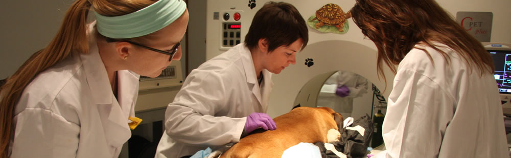
PET scans use a small amount of radioactive material and a special camera to identify abnormal tissues, including infected or cancerous tissue. Most often, the radioactive material is linked to sugar, and the sugar is taken up by active tissues. PET scan is most useful when it is combined with CT imaging to both identify the abnormally active tissue and see what that tissue looks like anatomically. Because active muscle will take up the sugar molecule, animals must be kept very quiet for a period of time before the test. Typically, they are kept sedated first and then fully anesthetized for the actual test.


Radiography (X-rays)

Plain film radiographs, or X-rays, allow us to visualize the inside of the body. These images do a great job showing bone and can also be used to examine soft tissues such as the lungs, liver and kidneys. Because radiographs produce a two-dimensional picture, it is necessary to obtain at least two separate views so overlapping structures can be seen well. Although radiographs are useful, they have distinct limitations. For this reason, other imaging modalities such as ultrasound are often used along with radiographs.


Technetium scan

Technitium-99m is the most widely used radioactive tracer in nuclear medical procedures. It is tagged, or bound to, a pharmaceutical that has an affinity for a particular tissue type. In veterinary medicine, bone scans and thyroid scans are the most common technetium scans. Patients must be housed in special radiation suites for 24 hours following injection of the radiotracer.


Ultrasound

Ultrasound uses sound waves to create images of the structures inside the body; many people are familiar with such imaging studies when performed on pregnant women to examine the growing fetus. We often use this technology to examine the contents of the abdomen and sometimes use it to look at other parts of the body as well. The ultrasound images are complementary to conventional radiographs (X-rays). In fact, it is routine to obtain radiographs prior to ultrasound exam so important findings are less likely to be missed.

In order for the sound waves to create a good image, the hair over the area is usually clipped, and a gel substance is placed on the skin. The procedure itself is not painful and is extremely safe, and it can often be completed with the animal awake. Sometimes sedation is required for pets that are anxious or squirmy. Although the ultrasound examination is not invasive, it is often used to help guide us to a specific site to collect biopsies or needle aspirates from diseased tissues. Sedation or even anesthesia may be required to make sample collection as safe as possible.


Oncology

  • About Cancer
    • Common Tumor Types
    • Cancer Effects
    • Common Cancer Terminology
  • Common Diagnostic Procedures
  • Cancer Treatment
    • Surgery
    • Chemotherapy
    • Radiation Therapy
  • Clinical Trials
    • Current Clinical Trials
    • Tissue Collection Bank
  • Your Appointment
    • Pet-Friendly Lodging
    • Flying with Pets
  • Client Support
  • Information for Veterinarians
  • Frequently Asked Questions
  • How You Can Help

Contact

Small Animal Hospital
573-882-7821; After Hours: 573-882-4589
Regular hours: M-F, 8 a.m. to 5 p.m.
Emergencies: Please see current policies


Equine Hospital
573-882-3513; After Hours: 573-882-4589
Regular hours: M-F, 8 a.m. to 5 p.m.
Emergencies: Open 24 hours a day


Food Animal Hospital
573-882-6857; After Hours: 573-882-4589
Regular hours: M-F, 8 a.m. to 5 p.m.
Emergencies: Open 24 hours a day

Our Location

900 E. Campus Drive, Columbia, MO Veterinary Medical Teaching Hospital

Giving to the VHC

The MU Veterinary Health Center is a leader in animal disease diagnosis, patient care and education. Individuals and corporations from all over the world are making a difference every day by providing support for our facilities, programs, students and faculty.

Learn how you can help us continue provide the most cutting-edge, compassionate care.
© 2025 — Curators of the University of Missouri. All rights reserved. DMCA and other copyright information.
An equal opportunity/access/affirmative action/pro-disabled and veteran employer. Disability Resources