The Orthopedic Surgery division of the Small Animal Surgical Service is focused on treating conditions involving the musculoskeletal system of your dog or cat including bones, joints, muscles, tendons and ligaments. Many such disorders do not require surgery, and can be managed medically. If surgery is required, however, it is possible that minimally invasive techniques may be employed to reduce surgical discomfort, shorten hospitalization and speed recovery.
The University of Missouri’s Small Animal Surgical Service was among the first in the world to employ such minimally invasive techniques including arthroscopy (exploration of the joint through tiny incisions using a camera) and fluoroscopic-guided fracture repair techniques in which large surgical approaches are avoided. We are proud to be a leader in the field in these techniques and to offer them to you.
Below is a short list of some of the more common conditions and surgical techniques we specialize in, categorized by the anatomic site affected. If you are interested in having one of our surgeons examine your pet for a suspected orthopedic problem, please contact us.
[accordion multiopen=”true”]
[toggle title=”Knee” state=”closed”]
Cranial Cruciate Ligament Disease
Many conditions can affect the knees of dogs and cats, but the most common disorder is tearing of the cranial cruciate ligament (CrCL), which is analogous to the anterior cruciate ligament (ACL) in people. ACL injuries in people almost exclusively occur secondary to trauma or excessive force applied to the knee. However, in dogs it is usually a degenerative process that doesn’t necessarily require a traumatic event. Once the ligament is torn, the dog will become pronouncedly lame on the affected limb and may not even be able to bear weight. This may improve slightly over time as the body tries to stabilize the joint, but a significant lameness usually persists. CrCL injuries can be managed medically, but typically dogs will not be able to fully recover without surgical stabilization because the CrCL is unable to heal once torn. Diagnosis of the condition is typically made during orthopedic examination and upon X-raying the knee.
There are many ways to surgically stabilize the knee once the CrCL has been damaged. Two of the more common procedures offered include the following:
Lateral fabello-tibial suture
Historically, this has been the simplest and most straightforward method of stabilizing the cruciate ligament-deficient knee. Although there is some variation in technique, the procedure essentially involves the passage of a suture from the back of the femur to the tibial crest in a direction oriented to mimic the absent CrCL. It is used more frequently in smaller dogs and cats. Advantages of the technique include lower risk of complication and expense as the procedure is less invasive than some of the alternatives. However, several recent publications have shown that this technique is less effective in providing comfort and return to function in medium and larger dogs over the long term than the next technique discussed, the tibial plateau leveling osteotomy, or TPLO.
Tibial Plateau Leveling Osteotomy (TPLO)
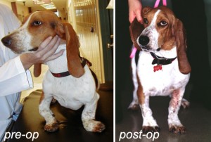
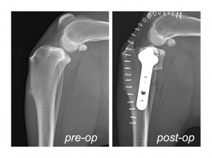 The TPLO technique is designed to alter the biomechanics of the knee such that the dog no longer requires the damaged CrCL to provide stability to the knee. This is accomplished by reorienting the surface of the tibia, which is the bone that makes up the bottom of the knee joint. Dogs possess a sloped proximal tibia, which can result in the femur sliding off the back of the tibia when they bear weight on their limb if the CrCL is damaged. The TPLO technique levels this slope, thereby preventing the dynamic instability of the joint. Several publications now suggest that this technique is superior in restoring comfort and function over the long term. The surgeons of the Small Animal Surgical Service employ the most advanced instrumentation available when executing this technique to reduce the risk of complication and optimize the outcome for your dog.
It’s important to remember that once the CrCL has been damaged, osteoarthritis begins to develop in the knee. No stabilization surgery can stop this process or reverse its effects. Thus, even though the prognosis for return to function and resolution of discomfort is very good with these stabilizing techniques, managing osteoarthritis will be a lifelong prospect.
Patellar Luxation
The patella, or knee cap, can sometimes become loose and displaced in dogs and cats, a condition we refer to as patellar luxation. If severe, this can result in pain and disability in the knee because when working properly, the patella serves to act as a pulley in redirecting and conducting the forces of the large quadriceps muscles of the thigh to extend the knee. If the patella is displaced, the animal can have difficulty using the knee appropriately and be painful. Diagnosis of this condition is made during the orthopedic examination and the completion of X-rays. A variety of surgical techniques can be employed to successfully correct this condition safely depending on the type and severity of the luxation.
[/toggle]
[toggle title=”Hip”]
Hip Dysplasia
Hip dysplasia is a multifactorial disease that is biphasic, meaning that it has a different presentation in very young dogs than it does in an adult dog. In puppies, it may present simply as looseness in the hips, but in older dogs will typically be accompanied by degeneration and remodeling of the bones that make up the ball-and-socket joint. Dogs with hip dysplasia usually exhibit lameness after a lot of activity and can be stiff in the morning. Many will show a classic “bunny-hopping” gait in the rear limbs while running. Because of this difference in presentations, different techniques are available to intervene depending largely on the age of the dog and the presenting signs, which are discernible on orthopedic examination and X-rays:
Puppies 12 – 18 weeks of age: If a puppy of this age is suspected to be at risk for canine hip dysplasia, a procedure called a juvenile pubic symphysiodesis (JPS) can be completed. This technique fuses a growth center of the pelvis, which improves the coverage of the ball of the femoral head by the socket of the pelvis, called the acetabulum. It can be done as an outpatient procedure during the same anesthetic episode as your dog’s spay or castration, and has a good chance of preventing hip dysplasia in a large number of dogs. Consult your veterinarian or make an appointment with us to determine if your dog is a candidate for a JPS.
Puppies 5 months – 12 months: Dogs of this age are too old to receive a JPS, but may be candidates for another procedure that also increases socket coverage of the femoral head through alteration of the bones of the pelvis in a technique called a double pelvic osteotomy (DPO). While more invasive than the JPS, this is still a very successful technique to help prevent the severe secondary changes that can result from hip looseness in young dogs. Consult your veterinarian or make an appointment with us to determine if your dog is a candidate for a DPO.
Dogs 11 months and older: Typically, dogs of this age are too old to be good candidates for a DPO because secondary remodeling changes have started to affect the dysplastic hip that was loose as a puppy. A variety of management protocols are possible for dogs in this category, including both non-surgical and surgical options. We can help you decide which is the best treatment for your dog depending on the severity of disease, clinical signs, the changes seen on X-rays and your dog’s lifestyle. There are usually several options that may work. If surgery is indicated, there are two basic choices:
Femoral head and neck excision (FHNE): The femoral head and neck excision is a technique by which the femoral head and neck (which comprise the “ball” of the ball-and-socket joint) are surgically removed to alleviate the bone-on-bone pain associated with advanced canine hip dysplasia. It can be completed in any size dog, but appears to work best in small dogs and cats whose hip disease is no longer manageable with medical techniques. Advantages of this technique are its low cost; however, it can result in some gait abnormalities and may not eliminate all discomfort in all dogs, especially larger ones.
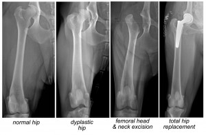 Total hip replacement (THR): Considered the gold standard in treatment of severe hip dysplasia that is no longer manageable with medical treatments, the total hip replacement involves the replacement of the affected femoral head and acetabular socket of the hip joint with artificial replacement components fashioned from medical cobalt-chrome and polyethylene. Advantages of this technique are the expected return of full function and resolution of pain for the dog for the rest of its life. Although more expensive and requiring a longer post-operative recovery time than FHNE, medium- and large-breed dogs typically have far superior function with this technique.
[/toggle]
[toggle title=”Elbow”]
Elbow Dysplasia
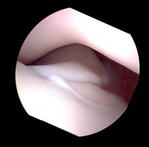 Elbow dysplasia is the most common cause of forelimb lameness in dogs. Like hip dysplasia, it is a multifactorial complex condition that will first affect the dog as a puppy. Over time, elbow dysplasia may result in loss of cartilage and remodeling of the joint surfaces with secondary osteoarthritic changes. The term elbow dysplasia actually describes a disease that comprises a number of different conditions. Through orthopedic examination and X-rays, the surgeons of the Small Animal Surgical Service will be able to discern what, if any, components of elbow dysplasia your dog possesses to help guide treatment decisions. Treatment will vary depending on which of these contributing conditions is present and can consist of medical management, arthroscopic examination (via a very small incision and the insertion of a tiny camera) of the joint, removal of cartilage and bone fragments or corrective osteotomies (cuts in the bone) to correct alignment. The recommended treatment will be tailored specifically to the needs of your dog.
[/toggle]
[toggle title=”Shoulder”]
Shoulder OCD
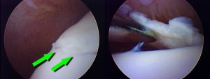 OCD stands for osteochondritis dissecans and describes a condition whereby a portion of the cartilage of the joint doesn’t form normally and can become dislodged, resulting in inflammation and pain. Much like elbow dysplasia, this condition will affect puppies and present as a forelimb lameness. The diagnosis is made upon orthopedic examination and taking X-rays of the shoulder joints. This condition is usually successfully treated via arthroscopic removal of the fragment. This is a minimally invasive procedure as it only requires two tiny incisions: one for the camera and one for a small instrument to retrieve the cartilage fragment and to clean up the joint. The prognosis for return to function and alleviation of pain is excellent.
Biceps Tenosynovitis
In very active and working dogs, the large tendon that spans the shoulder joint, called the biceps tendon, can become injured, resulting in an intermittent forelimb lameness. Once injured, it can be a source of inflammation and discomfort if not treated. The diagnosis of this injury is completed on orthopedic examination and diagnostic imaging, including X-rays and ultrasound. Depending on the chronicity, severity and type of injury, different treatment protocols can be employed, ranging from medical management to minimally invasive surgical intervention.
Shoulder Instability
The shoulder joint is considered a “ball-and-socket” joint and is stabilized by a number of different tendons, ligaments and the joint capsule. Occasionally, in middle-aged and older dogs, some of these tissues can become stretched or damaged resulting in instability of the joint. The diagnosis of this condition can be challenging and requires a thorough orthopedic examination and usually some kind of diagnostic imaging like ultrasound. Treatment will vary depending on the severity and chronicity of the condition, as well as the kinds of activities your dog partakes in. A treatment plan will be developed specifically for your pet based on these factors and may include physical rehabilitation, surgery or a combination of the two.
[/toggle]
[toggle title=”Bone”]
Fractures
Traumatic injuries resulting in bone fractures are common in dogs and cats. Some fractures can be managed with splints or casts. However, more commonly, surgery is indicated to quickly improve the comfort and mobility of your pet. The orthopedic surgeons of the University of Missouri’s Small Animal Surgical
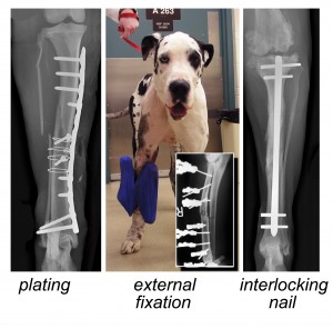 Service are skilled in a number of different fracture repair modalities including external fixation, plating and interlocking nails, and will employ the most practical, safe and effective techniques for your dog or cat. Frequently, these may involve what are called minimally invasive techniques, which require less aggressive surgical approaches that lessen the discomfort your pet experiences and quicken the rate of healing. The University of Missouri is a leader in the field of minimally invasive fracture repair techniques and is responsible for the development of many of these procedures.
Before your dog or cat is taken to surgery to repair the fracture, they will be fully assessed to determine if any other injuries were incurred during the traumatic event, and treated accordingly. With a full staff of criticalists, internists, radiologists and other specialists, we apply a team approach to the care of your pet to ensure that all of their needs are met.
Limb Deformities
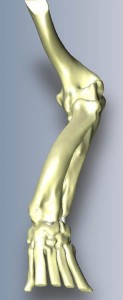 Limb deformities can affect both dogs and cats, causing shortening or angulation of a limb, resulting in disability and pain. These cases can be particularly challenging, but surgeons at the University’s Small Animal Surgical Service has focused a tremendous amount of research on this problem to become a leader in the field in the area of deformity correction. Frequently, deformities are first detected in the young animal, as the proper growth of the bone is affected. Therefore, surgical intervention in juvenile patients is often indicated. However, many times the deformities are not fully realized until adulthood, in which case different techniques are employed.
Deformities can be assessed with X-rays, but more frequently, we are moving toward computed tomography (CT) for their evaluation, which allows us to fashion 3D reconstructions of the limb for a more precise examination as well as the potential to complete rehearsal surgeries in advance of the actual procedure.
The Small Animal Surgical Service will work with you and your pet to develop a comprehensive treatment plan to correct the deformity and return your pet to a functional lifestyle.
[/toggle]
[/accordion]
If you have questions about any orthopedic condition in your dog or cat, or would like us to examine your pet and discuss treatment options, please contact us.

